
Arachnoid Cyst Information & Management
Introduction
An arachnoid cyst is a slow growing cyst found in the brain or spine, that contains clear fluid similar in composition to brain fluid or cerebrospinal fluid (CSF. Arachnoid cysts are congenital and benign cysts resulting from the splitting of the arachnoid layer of the meninges.
The brain and spinal cord is surrounded by a thick protective layer called the meninges. The meninges are attached to the inner aspect of the skull and contained within the spinal canal of the bony skeleton of the spine. The meninges consist of three distinct layers. The outermost thick layer is the dura, the middle layer is the arachnoid and the innermost layer, directly adjacent to the brain and blood vessels, is called the pia. Arachnoid cysts arise from the arachnoid layer.
Arachnoid cysts are often discovered incidentally and are usually classified depending on their location within in the skull cavity and size. Most arachnoid cysts do not require treatment. Rarely they can cause symptoms and require surgical intervention.
Why do patients develop arachnoid cysts?
Most arachnoid cysts are congenital lesions, meaning they are usually present from birth, although developments in adolescence have been described. Congenital cysts are termed primary arachnoid cysts. Rarely, secondary arachnoid cysts may develop later in life due to processes such as inflammation or trauma.
|
What are the signs and symptoms of arachnoid cysts?
Arachnoid cysts are usually asymptomatic. Usually they are discovered during investigations for other reasons, such as a CT scan of the brain performed following a traumatic head injury or for investigation of headache. Sometimes arachnoid cysts may cause symptoms related to pressure or mass effect within the brain. These symptoms include:
|
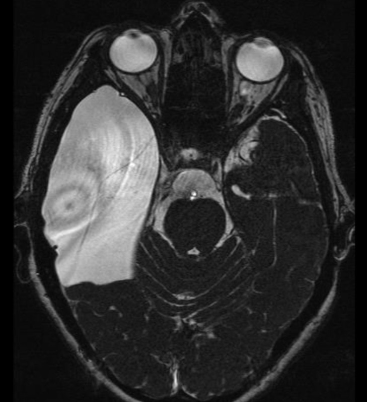
MRI scan of the brain demonstrating a large right temporal (middle fossa) arachnoid cyst. This patient presented with severe and progressive headaches with balance disturbance.
|
What investigations are performed for arachnoid cysts?
Routine imaging of the brain or spine is sufficient to identify most types of arachnoid cysts. Radiological investigations include:
What is the treatment of arachnoid cysts?
If an arachnoid cyst is discovered incidentally and is asymptomatic it does not need treatment. Cysts with the following characteristics can generally be watched and do not require surgical intervention:
If the cyst is causing headaches, raised intracranial pressure or focal neurological deficits, demonstrates significant growth, or appears atypical on MRI then neurosurgical intervention will sometimes be indicated.
Neurosurgical treatment options for arachnoid cysts include:
Surgical treatment of arachnoid cysts is generally well tolerated by patients and is relatively safe. Occasionally a second operation may be required, particularly if there is scarring at the previous operation site resulting in reformation/recurrence of the cyst. Shunting of the cyst is the last resort treatment option.
If you have recently been diagnosed with an arachnoid cyst please do not panic, these are very benign lesions that only very rarely require surgery. They are not cancerous. Dr Oehme will advise which treatment is most appropriate for your arachnoid cyst.
Routine imaging of the brain or spine is sufficient to identify most types of arachnoid cysts. Radiological investigations include:
- CT brain/head – an arachnoid cyst may be discovered incidentally following a scan of the head for another cause.
- MRI brain – an MRI gives higher definition of the cyst and is used to help plan surgical treatment if needed. It is also helpful in distinguishing between a benign arachnoid cyst and a cystic brain tumour. Once a cyst is discovered, MRI is usually performed to confirm that the cyst is, in fact, a benign arachnoid cyst. Other lesions which can mimic arachnoid cysts include cystic tumours, epidermoid lesions, Dandy Walker malformations or mega cisterna magna. Dr Oehme will be able to determine what type of cyst is present on the MRI scan and advise you on appropriate treatment accordingly.
What is the treatment of arachnoid cysts?
If an arachnoid cyst is discovered incidentally and is asymptomatic it does not need treatment. Cysts with the following characteristics can generally be watched and do not require surgical intervention:
- Cysts that are not causing symptoms
- Cysts that are small in size
- Cysts which do not show growth on repeat brain imaging
- Cysts that are not causing any pressure on the brain
- Cysts with MRI features classic of arachnoid cyst
If the cyst is causing headaches, raised intracranial pressure or focal neurological deficits, demonstrates significant growth, or appears atypical on MRI then neurosurgical intervention will sometimes be indicated.
Neurosurgical treatment options for arachnoid cysts include:
- Monitoring the cyst with serial imaging/MRI scans to detect for growth is the most common management option.
- Surgery to drain (aspirate) and/or excise the wall the cyst. This generally involves a craniotomy to access cysts in the brain, or a laminectomy to access a cyst in the spine. If possible the walls of the cyst are removed to allow free flow of
cyst fluid with the surrounding CSF. The biggest risk of simple drainage/aspiration of the cyst is recurrence of the cyst, meaning it can grow again. - Surgery to make the cyst communicate with other fluid spaces in the brain – known as fenestration of the arachnoid cyst. This generally involves a craniotomy to access the cyst and to then open it so that its fluid contents drains into the basal CSF cisterns. A minimally invasive endoscopic approach may be used in some instances rather than a craniotomy.
- Surgery to shunt fluid from the cyst to the lining of the abdomen - cysto-peritoneal shunt. This allows for the flow of cyst fluid into the abdominal cavity, thereby decompressing the cyst and relieving pressure on the brain. See our VP shunt page for more information about surgery to insert a cranial shunt. Shunting is typically reserved for recurrent arachnoid cysts and is rarely used as an initial treatment.
Surgical treatment of arachnoid cysts is generally well tolerated by patients and is relatively safe. Occasionally a second operation may be required, particularly if there is scarring at the previous operation site resulting in reformation/recurrence of the cyst. Shunting of the cyst is the last resort treatment option.
If you have recently been diagnosed with an arachnoid cyst please do not panic, these are very benign lesions that only very rarely require surgery. They are not cancerous. Dr Oehme will advise which treatment is most appropriate for your arachnoid cyst.